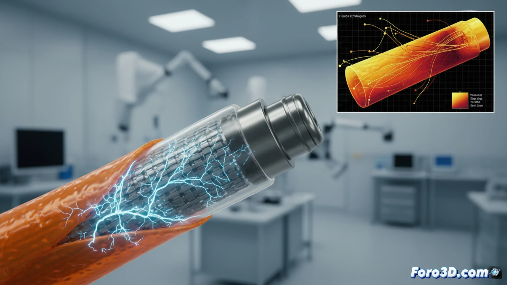
A patient undergoing cochlear implantation experienced sudden and irreversible hearing loss after device activation. Clinical suspicion pointed to a fatigue fracture in the platinum electrode array, induced during automated surgical insertion. To confirm the failure, a 3D expert analysis using micro-CT was employed, whose volumetric analysis revealed microcracks at the interface between the metal and the insulating polymer.
Forensic workflow: from tomography to finite element simulation 🛠️
The process began with the acquisition of micro-CT images of the explanted device. These were imported into Materialise Mimics to segment the actual geometry of the electrodes and the cochlea, generating a high-fidelity surface model. Subsequently, the mesh was transferred to Volume Graphics VGSTUDIO MAX for internal defect inspection and detection of submillimeter cracks. Finally, the cleaned model was taken to ANSYS for a Micro-FEA analysis, where cyclic loads equivalent to insertion forces were applied. The results showed a stress concentration at the electrode curvature, exceeding the fatigue limit of platinum after approximately 50 load cycles.
Lessons for critical implant design 💡
This case demonstrates that material fatigue simulation is not just a design tool but a cornerstone in clinical failure investigation. The integration of micro-CT with FEA allows validating fracture hypotheses that escape the human eye or conventional optical microscopy. For biomechanical engineers, the message is clear: any microcomponent subjected to cyclic stress during surgery must be modeled with the patient’s actual geometry. Without this workflow, the failure would have gone undiagnosed, perpetuating a risk in future insertions.
As a simulation engineer, what practical lessons can we draw from the correlation between micro-CT data and FEA results to predict fatigue failure in platinum electrodes before cochlear implant activation?
(PS: Material fatigue is like yours after 10 hours of simulation.)