Sonosensitizer performance
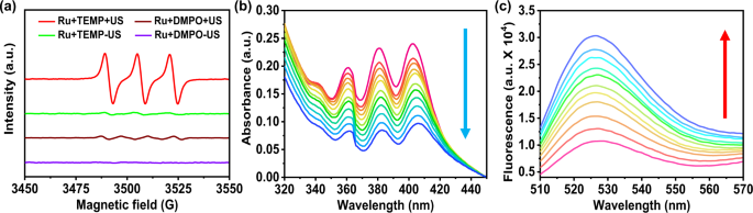
[Ru(bpy)3]2+ has great advantages for research application such as simple structure, easy synthesis, and high yield. The MS, UV-visible absorption, 1H NMR, 13C NMR and emission spectra of [Ru(bpy)3]2+ were characterized in the supporting information (Supplementary Fig. 1). To investigate whether [Ru(bpy)3]2+ could use for SDT, we firstly employed electron spin resonance (ESR) to detect the ROS generation. The spin trap 2,2,6,6-tetramethylpiperide (TEMP) as the 1O2 trapping agent. As shown in Fig. 1a, we can observe there 1:1:1 intensity signals appeared between 3480 and 3530 GM in TEMP and [Ru(bpy)3]2+ mixing solution under US irradiation. In contrast, the ESR spectra of water solvents alone by US irradiation were studied as Supplementary Fig. 2. We did not find obvious 1O2 and •OH generation in water solution under US irradiation. Thus, [Ru(bpy)3]2+ excited by radical products of sonolysis of water was not the dominant mechanism of [Ru(bpy)3]2+ excited by US.
![Fig. 1: Sonosensitizer performance of [Ru(bpy)3]2+.](https://media.springernature.com/lw685/springer-static/image/art%3A10.1038%2Fs41467-021-25303-1/MediaObjects/41467_2021_25303_Fig1_HTML.png?as=webp)
a ESR spectra demonstrating 1O2 generation of [Ru(bpy)3]2+ under US irradiation (0.3 W cm−2, 3 MHz, 1 h). The TEMP and DMPO were used as 1O2 and •OH trapping agents, respectively. b Time-dependent oxidation of DPA indicating 1O2 generation by [Ru(bpy)3]2+ under US irradiation (0.3 W cm−2, 3 MHz). c Time-dependent 1O2 generation of [Ru(bpy)3]2+ detected by fluorescence intensity of SOSG under US irradiation (0.3 W cm−2, 3 MHz). The colored lines represent spectra recorded every 10 min for 100 min in (a) and (c). Ru: [Ru(bpy)3]2+. TEMP: 2,2,6,6-tetramethylpiperide; DMPO: 5,5-dimethyl-1-pyrroline-N-oxide.
We further measured the quantum yield of 1O2 by the oxidation of 9, 10-diphenanthraquinone (DPA)41,42. In the presence of 1O2, DPA would be oxidized to 9,10-diphenanthraquinone dioxide (DPO2), which has no obvious absorption in visible light band. As shown in Fig. 1b, with the increasing of US irradiation time, the characteristic absorption of DPA decreased gradually. The absorption peak of DPA around 378 nm was collected to measure the rate constant for DPA oxidation, and we find DPA oxidation versus time revealed linear relationship with the calculated rate constant was 0.00142 min−1 (Supplementary Fig. 3a). However, the DPA oxidation by [Ru(bpy)3]2+ without US irradiation or US alone was very slightly (Supplementary Fig. 4). In addition, singlet oxygen sensor green (SOSG) was used to track the capture of 1O2, along with its unique maximum fluorescence intensity at 525 nm. As shown in Fig. 1c and Supplementary Fig. 3b, the SOSG fluorescence intensity strengthened by degrees when [Ru(bpy)3]2+ and US irradiation were applied.
In the other hand, for •OH trapping agent, 5,5-dimethyl-1-pyrroline-N-oxide (DMPO) was used instead of TEMP at the same conditions. The result showed no obvious •OH signal in [Ru(bpy)3]2+ sample under US irradiation (Fig. 1a). Moreover, methylene blue (MB) was used to track the capture of •OH43. From Supplementary Fig. 5, we could not observe any change of absorption curve of MB, which verified [Ru(bpy)3]2+ with US irradiation can hardly produce •OH. In addition, we investigated the sono-stability of [Ru(bpy)3]2+ after 1 h US irradiation by detecting its absorption spectra and emission spectra (Supplementary Fig. 6). The results showed that [Ru(bpy)3]2+ exhibited high sono-stability under US irradiation, suggesting that [Ru(bpy)3]2+ has the potential to be an excellent sono-sensitizer.
Sonocatalytic oxidation of NADH
NADH is an important coenzyme, which participates in over 400 intracellular redox reactions. The selective induction of NADH ruin the redox balance and exterminate cancer cells44. Therefore, we quantified the sonocatalytic oxidation of NADH by [Ru(bpy)3]2+ under US irradiation. With increasing time of US irradiation, the NADH (150 μM) characteristic absorption peak around 339 nm decreased obviously in the presence of 10 μM [Ru(bpy)3]2+ (Fig. 2a and Supplementary Fig. 7). The NADH oxidation turnover frequency (TOF) was 3.62 h−1 counted from the disparity in NADH consistence following US irradiation. The characteristic absorption peak of NADH around 339 nm was collected to measure the rate constant for NADH oxidation, and we found NADH depletion versus time showed a first-order kinetics relationship, and the calculated rate constant was 0.0385 min−1. Importantly, by adding NaN3 as a 1O2 scavenger, we found that the NADH depletion rate by [Ru(bpy)3]2+ based SDT was not affected (Supplementary Fig. 8), indicating that [Ru(bpy)3]2+ was a sonocatalyst.
![Fig. 2: Sonocatalytic oxidation of NADH by [Ru(bpy)3]2+ under US irradiation.](https://media.springernature.com/lw685/springer-static/image/art%3A10.1038%2Fs41467-021-25303-1/MediaObjects/41467_2021_25303_Fig2_HTML.png?as=webp)
a The oxidation of NADH (150 μM) by [Ru(bpy)3]2+ (10 μM) under US irradiation in PBS solution, as monitored by ultraviolet–visible spectroscopy. The direction of change in absorbance with time is indicated by the arrows. US irradiation time: 0, 20, 40, 60, 80, 100, 200 min. Insert: Plots of lnA/A0 at 339 nm against time. b ESR spectrum of NAD• radicals trapped by CYPMPO demonstrating NADH oxidized by [Ru(bpy)3]2+ under US irradiation. CYPMPO (1 mg) was used for NAD• radicals in 50 μL PBS solution containing [Ru(bpy)3]2+ (5 mM) and NADH (10 mM) under US (0.3 W cm−2, 3 MHz, 1 h) irradiation. c Sonocatalytic oxidation of NADH (1.1 mM) by [Ru(bpy)3]2+ (0.1 mM) in D2O/CD3OD (1:1, v/v) with US irradiation (0.3 W cm−2, 3 MHz, 20 min). Peaks labeled with red triangles represent NADH and peaks labeled with blue circles represent NAD+. d The proposed mechanism of 1O2 generation and NADH sonocatalytic oxidation by [Ru(bpy)3]2+ under US irradiation. Ru: [Ru(bpy)3]2+; TOF: turnover frequency; CYPMPO: 5-(2,2-dimethyl-1,3-propoxycyclo-phosphoryl)−5-methyl-1-pyrroline-N-oxide; NADH: 1,4-dihydronicotinamide adenine dinucleotide.
To further confirm the NADH sono-oxidation, ESR was employed to trap radical intermediates during US irradiation. 5-(2,2-dimethyl-1,3-propoxycyclo-phosphoryl)−5-methyl-1-pyrroline-N-oxide (CYPMPO) was selected as carbon-centred free radical scavenger to detect NAD•. As shown in Fig. 2b, the signal of CYPMPO-NAD was detected by ESR in water solution contain NADH and [Ru(bpy)3]2+ under US irradiation. The result proposed NADH was converted to NADH+• through single electron transfer mechanism by shifting an electron to [Ru(bpy)3]2+, which was excited by US. Then NADH+• easily deprotonated and turn into NAD• radical, which later change into NAD+ by intramolecular migration32.
1H NMR spectroscopy was further used to monitor the transformation between NADH and NAD+. After being irradiated with US, NADH was transformed into its oxidized form NAD+ in the presence of [Ru(bpy)3]2+ (Fig. 2c). Some new peaks of NAD+ at 8.31, 8.55, 8.99, 9.36, and 9.58 ppm were observed. In contrast, no new peaks of NAD+ in the NADH alone, Ru alone, Ru + NADH, and NADH + US control groups were found. On the other hand, we further investigated NADH depletion in 4T1 cells (Supplementary Fig. 9). Under US irradiation, the intracellular NADH concentration reduced after incubation with [Ru(bpy)3]2+, while only US irradiation or only [Ru(bpy)3]2+ incubation, the NADH levels were unaffected. The results confirmed that [Ru(bpy)3]2+ upon US irradiation could induce NADH oxidation. As all the above results, [Ru(bpy)3]2+ can generate 1O2 and induce sonocatalytic oxidation of NADH under US irradiation, and its probable mechanism is shown in Fig. 2d.
In Vitro SDT of [Ru(bpy)3]2+
To evaluate in vitro sonotherapy efficiency of [Ru(bpy)3]2+, the methyl thiazolyl tetrazolium (MTT) assay was used to measure cytotoxicity of 4T1 murine breast cancer cells. Without US irradiation, [Ru(bpy)3]2+ exhibited no cytotoxicity in high concentrations (IC50 > 160 μM) for 48 h incubation (Supplementary Fig. 10a). For SDT evaluation, 4T1 cells were incubated with various concentrations of [Ru(bpy)3]2+ (0–20 μM) for 4 h, followed by US irradiation for different time durations (0–25 min). The US power of 0.3 W cm−2 was selected due to the temperature increase of the solution under higher US power (>0.3 W cm−2). For example, US irradiation with the power of 0.4 W cm−2 showed obvious heating effect on aqueous solution, and the temperature was high enough to kill 4T1 tumor cells directly (Supplementary Fig. 11a–c). In addition, the 4T1 cells killing efficiency decrease under lower power (0.1 W cm−2 and 0.2 W cm−2) of US irradiation (Supplementary Fig. 10b). And we chose 4 h incubation time of [Ru(bpy)3]2+ because it was well uptake by cells after 4 h, as shown in Supplementary Fig. 12. With US irradiation, the 4T1 cells viabilities continuously decreased with increasing concentration of [Ru(bpy)3]2+ (Fig. 3a). The IC50 value was calculated as about 2.91 μM. The same phenomenon was also observed in another cytotoxicity experiment designed as the same [Ru(bpy)3]2+ concentration (10 μM) and different US irradiation time (Fig. 3b). Moreover, to intuitive display the sono-cytotoxicity of [Ru(bpy)3]2+ on 4T1 cells, the treated 4T1 cells were co-stained with calcein AM (AM) and propidium iodide (PI) (Fig. 3c). As expected, only [Ru(bpy)3]2+ incubation or only US irradiation showed strong AM signal and weak PI signal, which meant little damage to 4T1 tumor cells. But the cells in [Ru(bpy)3]2+ and US irradiation group showed faint green fluorescence from AM and intense red fluorescence from PI. These results proved that [Ru(bpy)3]2+ exhibited high sono-cytotoxicity toward tumor cells.

a The cell viabilities of 4T1 cells after incubation with different concentrations of [Ru(bpy)3]2+ in the presence or absence of US. b The cell viabilities of 4T1 cells treated with [Ru(bpy)3]2+ (10 μM) for varied US irradiation time. c Confocal images of 4T1 cells stained with calcein AM (green, live cells) and propidium iodide (red, dead cells) after different treatments. The experiment was repeated three times independently with similar results. d Confocal images of 4T1 cells stained with SOSG (green) after various treatments. Ru: [Ru(bpy)3]2+; US: 0.3 W cm−2, 3 MHz, 20 min; AM: calcein AM; PI: propidium iodide. All cell viability data was performed as duplicates of quadruplicate (n = 4 biologically independent samples). Error bars represent S.D. from the mean. Statistical significance was calculated with two-tailed Student’s t test (a) and (b) (***p < 0.001, **p < 0.01, or *p < 0.05).
ROS generation in cells
To investigate cellular ROS of [Ru(bpy)3]2+ for sonotherapy, 2,7-dichlorofluorescein diacetate (DCFH-DA) and SOSG staining assays were used to confirm the intracellular ROS levels, respectively (Fig. 3d and Supplementary Fig. 13). The control, [Ru(bpy)3]2+ alone and US irradiation alone groups exhibited weak SOSG and DCFH-DA signal. In contrast, [Ru(bpy)3]2+ and US treated group showed strong green fluorescence from SOSG or DCFH-DA. The SOSG signal of [Ru(bpy)3]2+ and US treated group was decreased when NaN3 (a 1O2 scavenger) is present. The cytotoxicity of [Ru(bpy)3]2+ for sonotherapy on 4T1 cells was partly inhibited by adding NaN3 (a 1O2 scavenger) (Fig. 3c). These results suggested a large amount of intracellular 1O2 was produced and then kill cancer cells. To investigate the kinds of intracellular ROS of [Ru(bpy)3]2+ for sonotherapy, dihydroethidium (DHE) and 3′-hydroxy-6′-(4-hydroxyphenoxy)spiro[2-benzofuran-3,9′-xanthene]-1-one (HPF) staining assays were used to capture superoxide anion (O2−•) and hydroxyl radical (•OH), respectively (Supplementary Fig. 14). No obvious DHE signal or HPF signal could be found in the 4T1 cells treated with [Ru(bpy)3]2+ and US irradiation. These results excluded the effects of SDT on O2−• and •OH.
ROS generation in deep-tissue
Differ from PDT limited by the tissue penetration of light, sonotherapy is a promising new approach for deep-tissue tumor treatment due to excellent energy transfer efficiency of US. To investigate the sonotherapy efficiency in deep-tissue, 4T1 tumor-bearing mice were i.t. injected with SOSG and [Ru(bpy)3]2+ mixing solution and irradiated by US on the other side of the mice, which was far from the tumor side. The 1O2 generation of tumor tissue was detect by an in vivo fluorescence imaging system (Fig. 4a). After US irradiation, the fluorescence signal of SOSG in the tumor tissue was obvious, suggesting that the [Ru(bpy)3]2+ could generated 1O2 in deep tissue by US irradiation. Furthermore, a piece of pork (>10 cm) was selected to simulate human tissue for SDT-activatable depths research. SOSG and [Ru(bpy)3]2 + mixing solution was injected at different distances (every 2 cm position) from the US probe into pork. As shown in Fig. 4b, after US irradiation, the 1O2 generation was detected even up to 10 cm away from the US probe.

a The fluorescence imaging of 4T1 tumor-bearing nude mice with i.t. injection of SOSG and [Ru(bpy)3]2+ for varied US irradiation durations. The tumor tissue position is pointed out by red circle. b The fluorescence imaging to investigate 1O2 generation in the presence of [Ru(bpy)3]2+ and SOSG in deep pork tissue (>10 cm) under 30 min US irradiation. SOSG and [Ru(bpy)3]2+ mixing solution was injected at different distances (every 2 cm position) from the US probe into pork. c The fluorescence imaging of DAPI (blue) and DCFH-DA (green) co-stained tumor slices collected from mice after different treatments. Ru: [Ru(bpy)3]2+; US: 0.3 W cm−2, 3 MHz.
To further confirm the generation of ROS in tumor tissue, 4T1 tumor-bearing mice were sacrificed 2 h post various treatments. Their 4T1 tumor tissues were gathered for frozen sections and then stained by DCFH-DA and 4′,6-diamidino-2-phenylindole (DAPI), then these 4T1 tumor slices were photographed using a laser scanning confocal microscopy (LSCM) (Fig. 4c). Tumor tissue in [Ru(bpy)3]2+ + US group demonstrated intense green fluorescence due to sufficient ROS generated by [Ru(bpy)3]2+ based SDT. In contrast, the tumor sliced in the control, US and [Ru(bpy)3]2+ alone groups exhibited very weak green fluorescence signal. All results certificate that [Ru(bpy)3]2+ can generate ROS in deep-tissue in vivo.
Sonodynamic therapy in Vivo
Encouraged by the high 1O2 generation and sonocatalytic oxidation of NADH by [Ru(bpy)3]2+, we further investigated antitumor efficacy in 4T1 tumor-bearing mice model. The mice were divided into four groups (5 mice per group): (1) Untreated; (2) [Ru(bpy)3]2+ alone (i.t. injection 0.5 mg kg−1); (3) US alone (0.3 W cm−2, 3 MHz, 20 min); (4) [Ru(bpy)3]2+ + US 0.1 (i.t. injection 0.5 mg kg−1; 0.1 W cm−2, 3 MHz, 20 min); (5) [Ru(bpy)3]2+ + US 0.2 (i.t. injection 0.5 mg kg−1; 0.2 W cm−2, 3 MHz, 20 min); (6) [Ru(bpy)3]2+ + US 0.3 (i.t. injection 0.5 mg kg−1; 0.3 W cm−2, 3 MHz, 20 min). After 4 h post i.t. injection of [Ru(bpy)3]2+, the tumors were exposed by US irradiation. US at this power intensity was no thermal effect to kill tumor cells (Supplementary Fig. 11d, e). After that, the tumors were monitored by digital caliper and their volumes were calculated by the formula: Volume = 0.5 * Length * Width2 (Fig. 5a). The tumor growth was remarkably suppressed in [Ru(bpy)3]2+ + US 0.3 group, while tumors in the untreated group, [Ru(bpy)3]2+ alone group and US alone group showed obvious growth (Fig. 5b, c). In addition, 0.3 W cm−2 US irradiation showed improved inhibitory effect on tumors growth than 0.1 W cm−2 and 0.2 W cm−2 US irradiation. At the end of experiment, the mice in different groups were sacrificed so that the tumors can be gathered to photograph and weigh (Fig. 5d, e). Among the four groups, the average tumor weight in [Ru(bpy)3]2+ + US 0.3 group was the least (Fig. 5e). To confirm the efficient SDT in the deep-tumor tissues, the 4T1 tumor was transplanted on the right side of mice, and the US probe was on the left side of mice during treatment. The US wave penetrated from left to right. The result was shown in Supplementary Fig. 15, confirming that the efficient SDT of [Ru(bpy)3]2+ can reach deep-tumor tissues.

a Schematic of the in vivo sonotherapy procedure in 4T1 tumor-bearing mice. Mice was irradiated by US (0.1, 0.2, 0.3 W cm−2, 3 MHz) for 20 min after 4 h i.t. injected with [Ru(bpy)3]2+ solution. Tumor sizes were monitored every two days for 14 days in total. b Tumor growth curves of mice after various treatments. Error bars were standard errors (±SD) based on five mice in each group. Statistical significance was calculated with two-tailed Student’s t test, p = 0.000092 (***p < 0.001, **p < 0.01, or *p < 0.05). c Representative images of mice at day 14 after various treatments. d Average tumor weights of mice at 14 day post various treatments as shown in (e). e Photos of tumors were collected from mice at 14 day after various treatments. Ru: [Ru(bpy)3]2+. Error bars were standard errors based on five mice in each group. Statistical significance was calculated with two-tailed Student’s t test, p = 0.00023 (***p < 0.001, **p < 0.01, or *p < 0.05).
Furthermore, the sonotherapy efficacy was evaluated by haematoxylin and eosin (H&E) staining assay and TdT-mediated dUTP nick-end labeling (TUNEL) assay (Fig. 6a). 24 h after various treatments, the mice were sacrificed to collect their tumors for histological analysis. Consistent with the above data, we observed severe tissue damage of the tumor tissue in [Ru(bpy)3]2+ and US treated group. In contrast, control, only [Ru(bpy)3]2+ or only US group showed no obviously tissue damage confirmed by both H&E and TUNEL staining slices. The tumor tissue in NaN3 and [Ru(bpy)3]2+ added with US irradiation group showed decrease tissue damage compared to [Ru(bpy)3]2+ and US treated group, but obviously tissue damage compared to the other three groups. The results suggested that SDT damage to tumor tissue was related to singlet oxygen generation (Fig. 6a). The biosafety of [Ru(bpy)3]2+ were evaluated by H&E stained slices of main organ collected from healthy mice i.v. injected with five times of the therapeutic dose (2.5 mg kg−1) of [Ru(bpy)3]2+ (Supplementary Fig. 16). The results showed that no obvious tissue damage was found from these slices. We further studied that the LD50 of [Ru(bpy)3]2+ was 3.89 mg kg−1 in the acute toxicity experiment. These results mean that [Ru(bpy)3]2+ is safe in vivo.

a Microscopy photos of H&E and TUNEL stained tumor slices. Tumors tissue were collected from mice at 24 h post various treatments. The experiment was repeated three times independently with similar results. b Photos of horizontal sections of India ink stained lungs were collected from mice at 40 day post various treatments. Spontaneous pulmonary breast cancer metastasis sites are pointed out by red circles. c Microscopy images of H&E stained lung slices collected from different groups of mice at 40 day post various treatments. The experiment was repeated three times independently with similar results. US irradiation: 0.3 W cm−2, 3 MHz; Ru: [Ru(bpy)3]2+.
Anti-metastasis to lung in vivo
Lung metastasis is usually discovered in advanced cancer, which result in a rapid death. 40 days following the mentioned four treatments, India-ink was tracheal injected to darken healthy alveoli and their 1 mm-thick horizontal sections of lung were collected to photograph. As shown in Fig. 6b, a large number of lung metastasis sites (the white tissue was circled) were observed in lungs collected from untreated, [Ru(bpy)3]2+ alone and US alone groups. In marked contrast, lung collected from [Ru(bpy)3]2+ + US group showed no metastasis site. Moreover, obvious tumor characteristic tissue (crowded cancer cells) could be found in the H&E stained lung slice collected from untreated, [Ru(bpy)3]2+ alone and US alone groups (Fig. 6c). These results indicated that [Ru(bpy)3]2+ based sonotherapy inhibited the progress of tumor lung metastasis.