
Crystallinity: X-ray diffraction
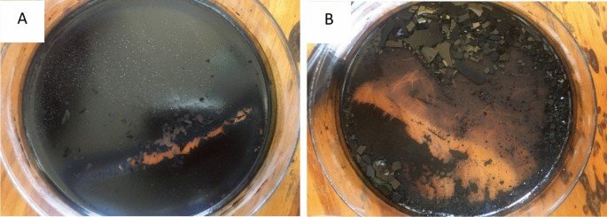
After the drying of ruthenium oxide nanoparticles, the blue-black crystals were also observed and shown in Fig. 1B. Figure 1A,B below shows the picture of ruthenium oxide nanoparticles in the process of drying. In this study, a Malvern PANalytical X’PERT PRO X-ray diffractometer with Cu Kα radiation of wavelength, λ = 0.15406 nm was used to determine the crystallinity of the powder prepared, its purity, and the structural phases in which it crystallized. XRD analysis (Fig. 2) showed the existence of crystalline RuO2 in a semi-amorphous state14, suggesting the formation of nanoparticles of RuO2. Bragg reflections at 33.18, 43.32, and 63.3° 2-theta with Miller indices (101), (210), (211) can be attributed to tetragonal RuO2 (JCPDS# 88-0322). The Bragg reflection observed at 16.49° 2-theta, we attribute to the presence of residual RuCl3/the formation of Ru-O-Cl15. The increase in background radiation observed at low 2-theta angles in the XRD pattern can be attributed to the scattering of radiation in the air, the geometry used for measurements, and some possible contributions from the sample which is in a semi-amorphous state.

(A) Shows the ruthenium oxide nanoparticles in the oven while drying, and (B) after drying.

X-ray diffraction of the semi-amorphous RuONPs obtained after drying the RuO-Gunnera perpensa extract at 60 °C for 24 h and 80 °C for 2 days.
The solubility of amorphous materials is much higher than that of crystalline16. An increase in solubility is significant as it can improve biopharmaceutical performance14,16. To estimate the average crystallite size of the RuONPs obtained after drying the Scherrer equation (Eq. 1) was used:
$$ {\text{D}} = K\lambda /\beta \cdot {\text{cos }}\left( \Theta \right), $$
(1)
wherein D is the particle size in nm, λ is the X-ray wavelength (Å), K is the shape factor, and Θ is half the Bragg angle, β is the Full Width at Half Maximum of the selected peak (in radians). Assuming a spherical shape (which is a generous approximation of the shape of RuONPs obtained in this work), with K = 0.94, and using the (210) peak the crystallite size was calculated to be 7.31 nm. The crystallite size of RuCl3/RuOCl impurity was calculated to be 0.24 nm.
Morphology: scanning electron microscopy
The structural morphology of synthesized ruthenium oxide nanoparticles was determined by Scanning Electron Microscopy. At a lower magnification of 10,000× (Fig. 3A), RuO nanoparticles were observed to be agglomerated. Higher magnifications at 60,000× in Fig. 3B showed that the ruthenium oxide nanoparticles were made of hexagonal-like platelets and giant octahedral cuboid-like structures at magnification. Agglomeration of the RuONPs is likely to result in a reduction in surface area and active sites available for interaction with cancer cells. Agglomeration coupled with the complexation of the Ru ions by biomolecules in the Gunnera perpensa plant extract may result in reduced activity of the RuONPs produced by this method of green synthesis. These two factors may account for the lower anti-cancer activity observed with the RuONPs when compared to Doxorubicin and Ru metal.

SEM micrographs show the morphology of ruthenium oxide nanoparticles at different magnifications (A) ×10,000; (B) ×60,000.
Absorbance: ultraviolet–visible spectroscopy (UV–VIS)
A Cary 5000 UV–VIS-NIR spectrophotometer was used to measure the absorbance of the synthesized RuONPs by dissolving the RuONPs in deionized water in a cuvette with a path length of 1 cm. Typically bulk RuO2 is metallic17. It should therefore absorb well into the Visible/IR region of the electromagnetic spectrum. Nanoparticles of RuO2 are reported in the literature to exhibit semiconductor properties and absorb between 1.0 and 2.2 eV (2.7 eV for RuO2 quantum dots)17,18. The absorbance peak observed (Fig. 4) shows that RuONPs prepared in this work absorb at the boundary of the UV/Vis region. Reduction of particle size from bulk to nanoscale (crystallite size = 7.31 nm) which results in the widening of the bandgap due to a quantum confinement effect would be observed as absorption in this UV/Vis region19. However the presence of residual RuCl3 in the RuONPs obtained after drying the RuO/Gunnera perpensa extract might contribute to the absorption. The predominance of the RuO2 in the sample is however made evident by the absorption at the boundary of the UV–Vis and the black colour.

Ruthenium oxide NPs UV–Vis spectrum showing the absorption edge at 413 nm. Inset (a) Gunnera perpensa extract in deionized water and (b) RuONPs (consisting of RuO2) dissolved in deionized water.
The profile of the UV–Vis absorption spectrum for RuONPs is akin to that reported by Anjum and co-workers19, in which they ascribe absorption at the UV–Vis boundary to RuO2 nanoparticles which they synthesized by annealing, at 600 °C, powders obtained from the plant-mediated reduction of RuCl3.xH2O using Moringa olifeira and Catharantus roseus. Based on the UV–Vis absorption spectrum in Fig. 4 and the assumption that the contribution of the RuCl3 impurity in the RuONPs is minimal the optical band gap (Eg) of what is predominantly RuO2 in a semi-amorphous state is estimated to be 3.00 eV using Eq. (2) where
$$ E_{{\text{g}}} = hc/\lambda , $$
(2)
and in which h = Planck’s constant, c = speed of light, λ = wavelength at the absorption edge. In terms of electron volts E (eV) = 1240 (eV.nm)/λ (nm) = 1240 eV.nm/413 nm = 3.0 eV. The value of 3.0 eV is not far off from the value of 2.7 eV reported by Parveen and coworkers for RuO2 quantum dots18. The contribution of residual RuCl3 to the calculated band gap of the RuONPs prepared in this work should however not be overlooked. The closeness of the value to that of RuO2 quantum dots19 (particle size = 3 nm) points however to the predominance of RuO2 in the RuONPs powder.
Chemical bond speciation using Fourier transform infrared spectroscopy (FTIR)
FT-IR spectroscopy was used to determine the chemical bonds present in the plant extract as well as those present in the prepared RuONPs. The IR absorptions for RuO2 have been calculated to be in the region of 466 cm−1, 669 cm−1, 1019 cm−1, 1648 cm−1 and 3408 cm−120. In comparison vibrations at 462, 1630 and 2889 cm−1 in Fig. 5B can therefore be attributed mainly to RuO2 whose unit cell is made up of 2 Ru atoms and 4 Oxygen atoms (Z = 2, space group P42/mnm), with a geometry of 4 long bonds Ru–O and 2 short Ru=O bonds21. Vibrations at 1338, 1584 cm−1 (due to in-plane CH2-bending), and 2889 cm−1 (CH2-bending and possible residual OH-stretching from waters of crystallization present in Ru hydrates) can be attributed to the interactions between RuO2 and residual organics still present in the RuONPs.
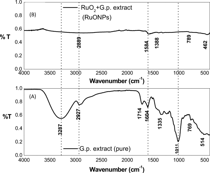
(A) FTIR spectrum of pure Gunnera perpensa extract after filtering and drying in air for 14 h (B) FTI-R spectrum of RuONPs after drying in air 60 and 80 °C for a period of 24 h and 2 days respectively.
In Fig. 5A the 514, 769, and 1101 cm−1 peaks depict C–H bending vibrations in phytochemicals present in the plant extract22. The peak at 2927 cm−1 can be ascribed to CH2– stretching vibrationsl11, while the strong broad peak at 3287 cm−1 points predominantly to the presence of H2O in the dried extract but also OH– stretching bonds found in the flavonoids, and steroids present23. The duplet at 1604 and 1714 cm−1 could be attributed to the presence of carbonyl groups (C=O) as may be found in steroids and flavonoids which are reportedly present in Gunnera perpensa.
It can be observed from Fig. 5A and B that peaks observed in the Gunnera perpensa extract are largely absent in the RuONPs—notes the almost complete disappearance of the 3287 cm−1 (H2O) 2927 cm−1 (CH2– stretching vibrations), a duplet at 1714 cm−1 (C=O), 1335 cm−1 (in-plane bending of CH2-groups), and 1011 cm−1. This suggests that: (1) much of the aqueous extract has effectively been separated from the RuO2 and any other Ru-complex formed both by decanting and drying off; (2) Ru metal and Ru ions in the Gunnera perpensa extract have effectively been complexed (chemically bound) to the phytochemicals present in the plant extracts; (3) the sample is quite dry (the strong broad peak at 3287 cm−1 observed in Fig. 5A due to water is almost completely absent in Fig. 5B).
Cytotoxicity activity of ruthenium oxide nanoparticles
Synthesizing nanoparticles from plant-derived materials for anticancer activity presents a promising paradigm shift in cancer treatment. The synergistic combination of plant compounds and nanotechnology has the potential to significantly enhance therapeutic outcomes, minimize side effects, and contribute to the development of more effective and patient-centered cancer therapies. The phenol, and flavonoids in the plant extract are converted to the corresponding aldehydes, carboxylic acids, ketones, and flavones during the plant-mediated production of metal nanoparticles, while the metal ions are reduced to form metal nanoparticles. Through the targeted delivery of medicines to cancer cells, Nano formulations can increase the effectiveness of Ru complexes in the treatment of cancer while minimizing side effects and systemic toxicity. Cell growth inhibitory activity of ruthenium oxide nanoparticles was evaluated on MCF7 (hormone receptor-positive breast cancer cell line) and doxorubicin was used as the standard drug for cytotoxicity and anti-cancer activity, respectively. Vero cells (kidney non-cancerous cell line) were used for selectivity.
Figure 6 shows that ruthenium oxide nanoparticles had the highest inhibitory activity of 22% at 10 µg/ml and the lowest inhibitory activity of 13% at 0.1 µg/ml. Our results showed that Vero cells and MCF7 cells viability were affected by synthesized ruthenium oxide nanoparticles, however, the inhibition was not selective. The percentage inhibition of doxorubicin on MCF7 ranges from 60 to 80% from a concentration range of 1 µg/ml to 100 µg/ml (as shown in Fig. 7).

Depicts the comparison of %Growth Inhibition of RuONPs vs the standard drug (Doxorubicin) against the Vero cell line. Tests were repeated in triplicates and data was represented as mean ± standard deviation.

Depicts the comparison of RuONPs vs Standard drug (Doxorubicin) against MCF7 cell line. Tests were repeated in triplicates and data was represented as mean ± standard deviation.
The results from this study show that the ruthenium oxide nanoparticle’s inhibitory activity against MCF7 cell lines was high when compared to the standard drug. The cytotoxicity of RuONPs was dose-dependent and the highest cytotoxicity was on MCF7 cells from the lowest concentration of 0.1 µg/ml. In addition, Fig. 7 shows the highest percentage growth inhibition of 59% as only 41% of cells were still viable after treatment with the standard drug at 0.1 µg/ml. while the lowest inhibition of more than 50% of the MCF7 cells was at 0.1 µg/ml for RuONPs and it was significant as compared to the control (p < 0.001). The IC50 value of RuONPs on both Vero and MCF7 cells was at 0.1 µg/ml, thus showing that RuONPs are highly cytotoxic to both Vero (non-cancerous) and MCF7 (breast cancer) cell lines.
A promising and novel anticancer drug should be selective in inhibiting cancer cells with minimal to no effect on the normal proliferation of non-cancerous cells. In this study, results show that RuONPs synthesized from Gunnera perpensa have a higher inhibitory activity on Vero cells, and thus could be considered cytotoxic to non-cancerous cells. Thus, suggesting that ruthenium oxide nanoparticles are not a good anti-cancer drug lead as they do not have selective cell inhibitory activity. According to Anjum et al.24, ruthenium as a compound has an anti-cancer activity, even though other studies showed the broad diversity of these compounds, in terms of activity, toxicity, and mechanisms of action25. Research findings by Liang et al.26, suggested that a compound (polypyridine ruthenium (II)) containing ruthenium has a high anticancer efficacy. Moreover, ruthenium (III) complexes have been reported to have a selective inhibitory effect on breast cancer cell lines and their mechanism of action is through the induction of apoptosis27. Contrary to this, ruthenium oxide nanoparticles synthesized from Gunnera perpensa did not show any selective activity. The isolated compounds from Gunnera perpensa have been reported to have antitumor activity on human breast cells in vitro28 and the crude extracts of this plant showed no cytotoxic activity on hepatic cells29. It should be noted that due to the fundamental nature of the synthesis process, existing approaches for making nanoparticles occasionally produce particles that lack selectivity30. The synthesis process may lead to the formation of non-selective nanoparticles with a wide size distribution and a variety of surface characteristics30. Thus, their efficacy in specific applications, such as medication delivery or cancer therapy, may be constrained by this non-selectivity. It should also be noted that there synthesized nanoparticles formed agglomeration as shown in Fig. 3. It is known that agglomeration/aggregation reduces the efficiency of nanoparticles and ultimately leads to subpar sample qualities31, thus, this could be another reason for the activity observed. From this study, it can be deduced that the combination of Gunnera perpensa and ruthenium oxide nanoparticles does not significantly enhance the anticancer activity of this plant as the ruthenium loses its selectivity.