
A new narrative review synthesizes the terminology, classification, plaque-related dosimetry, imaging biomarkers, and preventive and therapeutic evidence surrounding this condition, arguing that its biology is not a single linear sequence but an overlapping web of endothelial, inflammatory, permeability, ischemic, and neuronal changes.
The review, published in Frontiers in Oncology, was based on a PubMed/MEDLINE search covering January 2000 through 24 September 2026, using combinations of terms including uveal melanoma, radiation retinopathy, plaque brachytherapy, the radioisotopes iodine-125, ruthenium-106, palladium-103 and cesium-131, and imaging and treatment keywords such as OCT, OCT angiography, fluorescein angiography, laser speckle flowgraphy, anti-VEGF agents, corticosteroids and laser. Seminal pre-2000 studies that defined classification, natural history or treatment were retained, and original clinical studies were prioritized for efficacy and imaging claims while reviews supplied context.
A central message concerns structural optical coherence tomography. The authors note that OCT can reveal macular edema before ophthalmoscopic radiation retinopathy becomes visible, making it a sensitive early-detection tool. However, they caution that retinal thickness changes are not specific to edema: they can also reflect neuronal swelling in earlier phases and late atrophy, so thickness alone cannot be interpreted as a single disease stage.
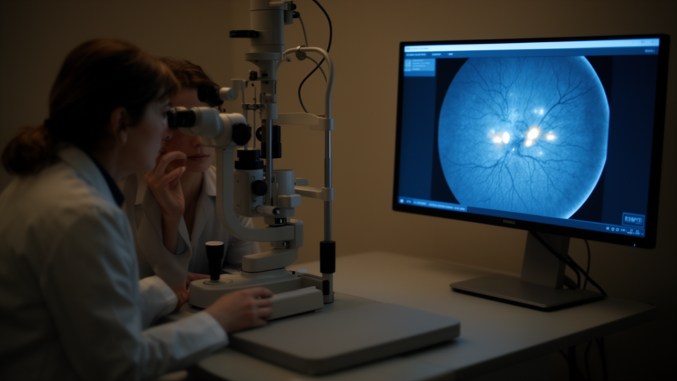
OCT angiography adds a perfusion dimension by detecting capillary-density loss and changes in the foveal avascular zone. Yet the review stresses that these measurements depend on signal quality and segmentation accuracy and are especially vulnerable to distortion in the presence of cystoid macular edema. Fluorescein angiography therefore remains important for assessing leakage and wide-field ischemic burden, while laser speckle flowgraphy is characterized as an investigational perfusion measure rather than an established clinical standard.
On treatment, anti-VEGF therapy emerges as the best-supported option for established radiation-related macular edema, but with an important caveat: it generally requires sustained dosing rather than short-course intervention. Prophylactic anti-VEGF cohorts show lower rates of edema and some other radiation complications, yet long-term visual superiority is not consistently demonstrated, and randomized, plaque-specific evidence is still lacking.
Corticosteroids and laser treatment retain selective roles in the management algorithm, according to the review, rather than serving as universal alternatives to anti-VEGF therapy.
The authors conclude that surveillance should be multimodal and individualized according to tumor location, patient-specific dosimetry, visual potential, and serial structural and perfusion findings. In their framing, OCT angiography complements rather than replaces structural OCT and angiography, and no validated OCTA threshold currently justifies initiating treatment in an otherwise unaffected eye.
Prospective studies, they argue, must now define reproducible biomarkers and treatment triggers so that early detection can be translated into evidence-based intervention. Until then, the review positions careful, individualized multimodal monitoring as the practical foundation for preserving vision in patients treated with plaque brachytherapy for uveal melanoma.
Subject of Research: Multimodal imaging biomarkers and management of radiation maculopathy after plaque brachytherapy for uveal melanoma
Article Title: Early detection and management of radiation maculopathy after plaque brachytherapy for uveal melanoma: a multimodal imaging narrative review
Article References: Gonzalez-Cortes, J. H., Gonzalez-Cantu, J. E., Folgar, A. M., Pelayes, D., & Mohamed-Hamsho, J. (2026). Early detection and management of radiation maculopathy after plaque brachytherapy for uveal melanoma: a multimodal imaging narrative review. Frontiers in Oncology, 16, Article 1926816. https://doi.org/10.3389/fonc.2026.1926816
Image Credits: AI Generated
DOI: 10.3389/fonc.2026.1926816
Keywords: radiation maculopathy, uveal melanoma, plaque brachytherapy, radiation retinopathy, optical coherence tomography, OCT angiography, fluorescein angiography, anti-VEGF, laser speckle flowgraphy, retinal ischemia, macular edema, Early