
Researchers at North Carolina State University have developed a molecular system that allows precise control over protein-degrading drug molecules using low-energy red light. The technology, termed RuPHOTACs or Ruthenium-based PHOToActivated Chimeras, keeps these molecules inactive until they are exposed to specific light conditions. This approach addresses a significant limitation in current drug therapies, where protein-degrading agents often affect healthy tissues alongside disease-associated cells. By controlling when and where these molecules become active, the system could help scientists develop treatments that destroy specific proteins while minimizing unwanted side effects in surrounding healthy tissue.
The study, published in Nature Communications, represents a proof-of-concept for using light to regulate PROteolysis-TArgeting Chimeras, or PROTACs. PROTACs are bifunctional molecules that work by simultaneously binding to a protein of interest and an enzyme that tags that protein for degradation. This process utilizes the cell’s natural waste removal mechanisms to eliminate the target protein. Unlike traditional inhibitors that block protein function, PROTACs catalyze the actual destruction of the protein, leading to a sustained and potent effect. However, the widespread presence of these target proteins in healthy cells poses a challenge for selective drug delivery.
Edith C. Glazer, a professor of chemistry at North Carolina State University and the corresponding author of the study, explained that most disease-associated proteins also perform important functions in healthy tissues. This dual role makes it difficult to target cancer or other diseases without causing collateral damage. Glazer noted that there are very few aberrant proteins found exclusively in cancer cells. Therefore, a method to control the activation of PROTACs is necessary to ensure they only function in the intended areas. The RuPHOTAC system provides this control by acting as a molecular cage that prevents the PROTAC from interacting with its targets until triggered.
The core of the RuPHOTAC system is a ruthenium complex, a metal from the platinum group of the periodic table. Ruthenium has specific properties that make it useful in light-driven chemical reactions. The researchers carefully designed this ruthenium complex to respond to relatively low-energy red light. This prevents the protein degradation process from initiating prematurely.
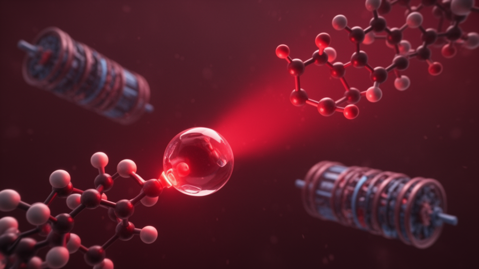
Glazer described the mechanism using an analogy, comparing the ruthenium complex to a beach ball that blocks two proteins from approaching each other. When the system is exposed to low-energy red light, the ruthenium is removed or released. The light-activated release provides a spatiotemporal control mechanism, ensuring that protein degradation occurs only at the specific location and time where the light is applied. This precision is crucial for reducing off-target effects in complex biological systems.
The researchers validated the RuPHOTAC system by targeting bromodomain-containing proteins, which act as crucial epigenetic regulators. They also demonstrated the system’s ability to strongly reduce levels of c-MYC and PIM1, proteins associated with various diseases. The study included both in vitro and in vivo testing, with the in vivo experiments conducted in a zebrafish model. The use of zebrafish allowed the researchers to observe the effects of the light-activated degradation in a living organism. The results showed that the RuPHOTAC was non-toxic in the fish model and effectively decreased the numbers of targeted proteins in specific areas when exposed to red light.
A key innovation in the study was the development of a new approach to monitor successful protein destruction. The researchers implemented a strategy involving the creation of a fusion between a photoconvertible protein called Dendra2 and the proteins of interest. This bifunctional reporter system allows for live cell analysis by decoupling protein degradation efficiency and rates from signals arising from new protein production. Traditional reporter systems often struggle to distinguish between the degradation of existing proteins and the synthesis of new ones. The Dendra2 fusion system directly measures the true degradation rate of the target protein in intact, viable cells while simultaneously tracking newly synthesized protein without perturbing translation, proteostasis, or cell health.
The study highlights several advantages of incorporating metal components within organic PROTACs for light-controlled systems. The RuPHOTACs demonstrated the highest selectivity for activation in the light versus the dark compared to other systems. This selectivity is critical for ensuring that the drug remains inactive until the precise moment of light exposure. The system also showed improved potency against the target proteins of interest. These properties make RuPHOTACs a promising tool for chemical biology applications where precise control over protein degradation is required.
Despite the promising results, the researchers noted limitations in the current system. The study is described as a proof-of-concept, indicating that further development is needed to optimize the system for broader biological applications. Glazer expressed hope that this work will lead to finer control over PROTAC technology. The ability to turn on protein degradation precisely where and when it is needed could address one of the major challenges in developing targeted medicines for various diseases.
The research was supported by the Kentucky Cabinet for Health and Family Services under Grant 1000200168, which provided the majority of the financial support. The National Institutes of Health also contributed to the project under grant R01GM162746, representing approximately 10% of the overall funding. The team included Dmytro Havrylyuk, Ainsley P. LaMore, David K. Heidary, and Edith C. Glazer from North Carolina State University. Majd A. Al-Hamaly and Jessica S. Blackburn from the University of Kentucky also contributed to the work. The authors declared no competing interests in the study.
Subject of Research: Development of ruthenium-based light-activated cages for controlling PROTAC-mediated protein degradation
Article Title: Researchers create light-activated cages for ‘protein killer’ molecules
Article References: Researchers create light-activated cages for ‘protein killer’ molecules. Original publication (Bibliographic details unavailable.)
Image Credits: AI Generated
DOI: Not provided
Keywords: RuPHOTACs, PROTACs, protein degradation, ruthenium complex, red light activation, North Carolina State University, Nature Communications, zebrafish model, Dendra2 reporter, bromodomain proteins, c-MYC, PIM1, spatiotemporal control, chemical biology, Edith C. Glazer, Dmytro Havrylyuk, Jessica S. Blackburn, University of Kentucky, drug targeting, epigenetic regulators